

Generally speaking, such nosologies as hidden caries are not in medical reference books and encyclopedias. Although dentists themselves and their patients actively use this term.
The point here is that hidden caries is the everyday name for any caries that cannot be found on teeth without special diagnostic methods. This is its main difference from caries in other manifestations: At the same time, the teeth look as absolutely healthy, but the pathological process in them can be so developed that for the treatment of the disease it will be necessary not only to remove part of the dentin, but also to depulp (“nerve removal”).
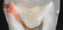
The photo below shows the hidden caries in the extracted tooth (located on the contact surface, that is, at the point of contact with the adjacent tooth). The lesion reached the pulp chamber:

In many cases, latent caries accompanies the usual and multiple, and only in exceptional cases does it develop independently among other healthy ones.teeth. Such cases are most rarely diagnosed and most often lead to very deep damage to the tooth.
Tooth lesions with hidden caries
In general, the damage to the teeth in the form of a cavity with hidden caries differs little from those for cases that can be distinguished by the naked eye. The nature and extent of such damage is determined by the age of the carious process and its localization on the tooth. So, hidden caries can have the following localization:
- On the proximal (contiguous) and distal (posterior) walls of the tooth. Here its manifestations on enamel are very difficult to notice with the help of a mirror or a probe.
- Under improperly placed seals or crowns. If bacteria from the oral cavity can penetrate the space between the tooth tissues and the filling material, the carious process is likely to develop here.
- In the areas of the tooth located under the gum (here, food particles often accumulate and are not cleaned with a toothbrush).
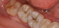
- In the fissures, natural fossae "rear teeth". If the damage to the enamel here is small, it is not always possible to notice it with the help of a mirror and a probe, and therefore even with the actual presence of external manifestations such caries can be attributed to hidden.
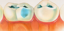
The photo is typical fissure caries with minimal external manifestations:

Damage to the tooth with hidden caries are similar to those with a visually diagnosed form of the disease. However, the specificity of the process leads to the fact that latent caries is usually found only in the middle and late stages of development, when they cover a large part of the dentin and parapulpal tissues. Accordingly, when the affected tooth is opened, extensive damaged cavities are found.
The following photos show an example of hidden caries in the tooth under the filling:


And in this picture - a radiograph of a tooth with hidden caries at a fairly late stage of development. It can be seen that the area affected by caries came close to the nerve canal:

From the practice of the dentist
A typical clinical case: in a patient among two teeth affected by caries, one seems to be healthy, with no visible damage. Logic dictates that if two teeth are affected, it is likely to be covered by caries and between them. I prescribe a radiograph to the patient in order to check the integrity of the internal tissues. On the radiograph notice well-developed caries near the posterior wall of the tooth.The lesion has not yet reached the pulp, but has penetrated deep into the dentin. In this situation, it was enough to clean out the damaged area and fillings.
Clinical manifestations
Hidden caries practically does not manifest itself. Since there is either no visible damage to the tooth enamel in it or it is small, the patient usually does not experience any unpleasant sensations when hot or cold fluids come into contact with the tooth, and normally chews it before it is involved in the pulp pathological process.
In rare cases, the affected area expands toward the adjacent areas of enamel and dark stains can be seen on a close look at the tooth. The photo below shows an example of such hidden caries under the old filling:

When the pathology spreads to the innervated areas of the tooth, the patient begins to experience pain when the tooth is exposed to cold or hot, as well as sour, sweet or salty foods. The first few weeks or months this pain is not very strong and does not always appear. Gradually, it increases and reaches a stagewhere the patient is forced to go to the clinic. In such situations, the depulpation is almost unavoidable (“nerve removal”).
What is dangerous hidden caries?
The main danger of hidden caries lies in the imperceptible development of the pathological process to such stages, which require either depulpation or tooth extraction in general.

In addition, with the development of caries on the proximal surfaces, its “transition” to the adjacent tooth is possible, and sometimes with the development and on it of the disease in a latent form.
On a note
It requires clarification: for some reason, many people are sure that caries can be “transmitted” and infect the adjacent tooth, but this is not so, caries is a local process and not “infectious”. It is important to understand that there will be no transition in the truest sense of the word to another tooth. Perhaps there is another thing: if a hidden caries has developed on one tooth with the formation of a cavity, then the self-cleaning of the gap leaves much to be desired. The constant accumulation of carbohydrate food remains causes the process to get worse in the first tooth and the beginning of a new destruction in the next.

In rare cases, or without pain, or with the patient's strong reluctance to consult a doctor, the carious area can cover most of the space filled with dentin.This situation is fraught with the fact that the tooth, in fact, turns into an empty shell, which can crumble even with a small load.
The photo below shows a hidden caries with a large lesion area (radiograph), the pathology covers a significant part of the tooth crown:

Diagnosis of the disease
Modern diagnostic methods allow to detect hidden caries at any of its localization and at any stage of development. But in order for it to come to the use of these methods, the dentist should suspect a carious process, even if it is indirect. Often, in the absence of patient complaints and when examining the oral cavity, hidden caries goes unnoticed and develops safely until pain in the affected tooth appears.
In many cases, latent pathology develops in parallel with carious lesions, which are clearly visible on other teeth. An experienced dentist in this case correctly assesses the overall cariogenic situation in the oral cavity, realizes that with frequent and multiple lesions of other teeth, the likelihood of hidden caries is very high, and prescribe diagnostics using special methods. These include, for example:
- A radiograph on which areas with hidden caries look darker compared to healthy neighbors.
- Transillumination and fluorescent diagnostics. Both of these methods reveal hidden carious tooth damage at any stage.
- Laser diagnosis.


It is almost impossible to find hidden caries on your own. Until the development of pulpitis, it will not be detected when a tooth is tapped and processed with cold or hot water.
Therefore, for early diagnostics of latent caries regular preventive visits to the dentist are very important.
How are hidden caries treated?
The treatment of latent caries in most cases consists in removing the affected cavities and further sealing the tooth. Moreover, if the pathology develops in the cervical area or under the gum, the dentist has to adjust the soft tissues surrounding the tooth.
The pictures below show examples of the preparation of carious cavities:


In some cases, hidden caries is found at the stage of stains or superficial enamel damage without penetration into the dentin.In these cases, for successful treatment it is possible to do without opening the tooth. The doctor can strengthen the enamel with calcium and fluoride to restore (remineralization).
In cases where the hidden carious process is found under the loosened seal, the damaged areas are cleaned and a new seal is installed in their place.
Such treatment is usually painless and can sometimes cause tingling. If a patient experiences acute pain when cleaning dentin, this indicates involvement of the pulp in the process and requires removal of the nerve with further filling of the canals.


In most cases, the treatment of latent caries with an operative method is carried out using anesthesia (local anesthesia), even if “drilling” is required not deeply. Usually this issue is consistent with the patient before treatment, and the decision is made mutually.
In rare cases, when a large-scale lesion of the tooth requires the excision of all tissues damaged by caries, up to the level of the gums, followed by the installation of a crown, or even tooth extraction and implantation of the prosthesis. As a rule, with regular visits to the doctor and regular check-ups, it is possible to avoid illness at such advanced stages and to preserve, though the filled, but native teeth.
Prevention of latent caries does not differ from that in its other forms: it is necessary to observe oral hygiene, remove food debris stuck between the teeth, limit the use of sweets in between meals, and use calcium and fluoride for prescribing by a doctor. And most importantly - regularly undergo examinations at the dentist. Hidden caries is a good example of the fact that a visit to the doctor is necessary even without pain in the teeth and other obvious signs of the development of the pathological process.
Causes of caries and effective methods of protection against it
And this is, in fact, the preparation of the cavity and the filling
The term "internal caries" ordinary patient dental clinic usually understands the disease, affecting tissue deep under the tooth enamel. ...
Decompensated form of caries is, in fact, an intensively developing caries, a pathological process that proceeds very actively in solid ...
Simplicity or, on the contrary, the complexity of diagnosing caries directly depend on its localization on the teeth and the stage of development. Hence, from the stage, ...
Thanks for the article, fully disclosed topic. As a patient, I was looking for a way to clarify secondary and latent caries. This is an x-ray. Thank you)) Good luck!
My doctor did not notice any hidden caries during the routine examination. After 6 months, the tooth began to crumble, and it turned out that caries was such that you need to put a crown ... That's how I lost a tooth. And, by the way, the doctor is very professional and expensive, from a private clinic.
And the doctor told me that there is no hidden caries on the X-ray + a nerve was removed in that tooth, and he said that it could ache gum. But more than a week has passed, and the tooth still hurts from below and between. What to do in this case?
Hello! The situation is difficult to remotely search for the cause of pain. There can be anything: from a mistake of a doctor in diagnosis to a banal inattention. I suggest one of the reasons:
one.Poorly treated tooth canals;
2. Hidden caries on the next "live" tooth;
3. Incorrectly formed contact point (gap between teeth), due to which the gum is injured.
You can send your snapshot of the tooth to the mail of the site - I will look and comment on it. With the help of images, the amount of information on a clinical case is often significantly increased. Be that as it may, your situation requires more careful consideration in order to exclude periodontitis in the tooth already treated in the canals, as well as possible pulpitis (or caries) in one of the neighboring ones.
Hello, a tooth with nerves hurts and gives down to the jaw. There was a filling, 5 years ago there was a caries, I treated it, after treatment for about a week the tooth ached, the doctor said that everything would pass - the pain passed. But over time, he began to get sick again, went to the doctor - he sent for an x-ray, said that everything was fine and he could not disturb, but replaced the seal (in my opinion, there was caries under it). But the tooth still hurts, and now the pain has intensified. What could it be? Thanks for the answer.