

Simplicity or, on the contrary, the complexity of diagnosing caries directly depend on its localization on the teeth and the stage of development. Consequently, the diagnosis of caries also depends on the stage at which the disease is located.
Thus, at the stain stage, the carious process is sometimes barely noticeable and can only be recognized by special means.


And already middle and deep caries can be recognized with the help of a standard dental mirror and probe. Moreover, the diagnosis is quite possible at home: multiple caries itself is conspicuous during the inspection of the oral cavity in the mirror, and even at the stage of the spot (initial caries) causes unpleasant sensations when hot or cold products hit the tooth, and sometimes even when cold air is inhaled.

However, caries is quite cunning, and in some cases it can develop asymptomatically to stages that require the removal of a significant part of the dentin, and sometimes depulpation (nerve removal). That is why the definition of pathology in dental clinics is often carried out with the help of modern equipment, and deep hidden lesions in the teeth are detected using high-tech methods.
Visual diagnosis of caries and cariogenic situation
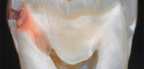
Visual inspection of the teeth is the main way to identify the cariogenic situation in the oral cavity. Caries is characterized by the fact that at almost all stages of its course it changes the color of the tooth enamel. Even at the stage of the stain, when dentin is not yet affected, the enamel is no longer smooth and shiny, and a careful dentist easily notices this change.

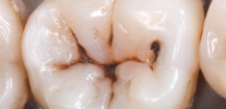
At later stages, it is even easier to identify caries during a simple examination: it either leads to the appearance of black and dark brown spots on the teeth, or if the dentin is damaged, dark cavities appear under the enamel.

Not surprisingly, in most cases, the visual diagnosis of dental caries allows you to identify most of the affected areas. With her, the dentist carefully examines the teeth from different sides with the help of a mirror. In addition, the doctor can probe the teeth - in the places of early development of the carious process, the enamel surface roughness is clearly felt.
On a note
It is visual diagnostics that is most accessible at home and makes it possible to recognize the pathology on the teeth independently. It is enough just to carefully inspect your teeth in the mirror. In this case, it is worthwhile not only to look for frankly black areas, but also to pay attention to all the places that stand out against the background of pure white teeth. Remember: black and brown areas indicate a fairly deep damage to the tooth (at best, only enamel will be pigmented). Such a tooth will probably have to be drilled, perhaps even a nerve will be removed from it. Therefore, a dentist should be contacted before significant visible damage occurs.
There are cases when visual diagnosis is not enough. For example, the affected areas that are on the wall of the teeth in the places of their contact are not always visible. Sometimes the carious process develops under old fillings and crowns, under the gum (root caries), and cannot be seen with the naked eye. Here, more accurate diagnostic methods come to the aid of the dentist.
Tooth staining, or the use of caries markers
The principle of caries-markers is that some substances linger precisely on the areas affected by caries.The most common such marker is methylene blue. When it enters the tooth from the healthy surface of enamel, it simply flows, and on carious, simply speaking, remains (diffuses into the pores of demineralized enamel). As a result, the doctor can not only determine dental caries, but also reliably identify the shape and extent of damage.

In most modern caries markers (they are also sometimes called caries detectors or indicators), magenta is used to color the affected areas pink. Markers are applied to the teeth with special tampons, and then washed off. The clinics use both imported products (VOCO, Pulpdent) and domestic preparations like the Color-test.
Roentgenogram of teeth
This method of diagnosing dental caries is especially good in situations where it is necessary to identify deep caries that have no noticeable external manifestations. For example, if the place of violation of the integrity of the enamel is hidden by the wall of another tooth or gum. On the radiograph, all internal cavities are well recognized, but in most cases it does not allow recognizing caries in the early stages of development.

The decision on the appointment of X-rays is always taken only by a doctor, and not all clinics have the appropriate devices. Usually they are quite large and expensive, and small cabinets do not have the opportunity to purchase even the simplest device. Patients of such cabinets are diagnosed in third-party clinics. It is clear that only for the diagnosis of caries such devices are not purchased - their functionality is much wider.
Thermodiagnosis caries
Diagnosis of caries on the teeth using low and high temperatures is usually carried out to detect damaged areas on proximal surfaces. At the same time, the tooth is simply treated with cold or hot (up to 60 ° C) water, and due to the patient's painful sensations or their absence, they conclude that there are carious lesions.

Usually, thermodiagnostics is used as an auxiliary method for confirming a previously established diagnosis. This method is also used to diagnose caries: if the pain passes quickly after contact with water, this indicates the presence of caries only. If the pain persists for a long time and is more acute, then we can already talk about pulpitis.
Electrodontometry
Doctors also do not often use electroodontometry (EOM) to diagnose caries. Basically, this method is used to detect pulp lesions, and therefore it is used to make a differential diagnosis of caries for pulpitis.

The principle of an EOM is to exert a different force on the pulp. Depending on the kind of current that causes pain, the doctor concludes that the pulp is involved in the pathological process.
On a note
Do not be afraid of electrical monitoring. Exposure to current does not mean that the tooth will be “hit” by electrical discharges. Usually the effect is stopped when the first tingles appear. This is enough for a doctor to determine the pathology. If the tooth has only caries, the EOM does not cause pain.
Fiber-optic transillumination (transluminescence)
Transluminescence and luminescent diagnostics close to each other are methods of caries diagnostics that are rarely used in medical practice. Usually they are used for other purposes.
- Fiber-optic transillumination is based on the illumination of the tooth with bright light.At the same time, the areas affected by caries turn out to be darker and create a clearly visible hemisphere against the background of healthy enamel. Such an examination is carried out in a dark room and use cold light.
- Fluorescent diagnostics is to illuminate the tooth with ultraviolet light. Usually a beam of light is passed through a special filter (Wood filter). In such a light, the tongue appears orange, healthy teeth have a snowy white shade, and carious - darker. The boundaries of the affected areas are very well marked and therefore easily recognized.


Both of these procedures are completely painless. Apparatuses for diagnosing caries by this method are produced by the domestic industry (OLD-41) and many foreign manufacturers.
There is also a method of laser-induced fluorescence. In it, the tooth is illuminated by a laser beam, and a conclusion is drawn about the integrity of the tooth enamel from the spectrum of its own radiation. Devices for such a diagnosis are small and are a good help in studying the cariogenic situation in the oral cavity.
Fissurotomy diagnosis of caries
Fissurotomy consists in opening the enamel in the area of dental fissures to identify areas damaged by caries under it. Of course, in standard situations such a procedure is not performed for diagnosis: it is impossible to just drill a tooth in order to make sure that it is healthy. As a rule, this method is used to confirm the physician’s suspicion of caries, as well as to assess the extent of damage of a uniquely carious tooth.

In general, this method is mainly the assessment of the lesion, but not the identification of the caries itself.
From dental practice
Often in patients on a particular tooth, a contact wall with gray enamel is found. Although caries is not clearly visible, under the most intact enamel an unknown formation is visible. In all cases, after treatment with fissure or simple spherical boron, the enamel falls through and a hidden vast cavity in the tooth opens. Very often, a chronic pulpitis develops under this seemingly intact wall.
Differential diagnosis: with what diseases of the teeth can dental caries be confused?
Differential diagnosis of dental caries is carried out with almost all the methods described above.Wherein:
- Staining caries-markers can distinguish caries from hypoplasia and fluorosis. Only caries will remain marked after applying the detector and rinsing it with water.

- EOM and thermodiagnostic distinguish caries from pulpitis and periodontitis.
- Based on an X-ray, deep caries can be distinguished from non-carious lesions of enamel and complications of caries (pulpitis and periodontitis).
Differential diagnostics are almost impossible to use with visual methods. Therefore, for example, at home to distinguish deep caries from pulpitis is possible only by the sensations and nature of the pain.
In general, it is possible to independently diagnose caries in oneself by noticing any abnormalities in the natural pure-white color of the tooth enamel. Especially carefully should inspect the areas around fillings, chips, over the gums. It is also necessary to pay attention to the sensitivity of the teeth. Sometimes enamel is chipped on the front teeth, leading to pain when breathing. Places of such chips are the "gateway" for the destruction of open areas of cariogenic bacteria.

An additional, though not very unambiguous method of self-diagnosis of caries may be the use of dental floss.With their help, you can find damage to the enamel, even on those sides of the teeth where caries is not noticeable. You just need to rub the thread of this side of the tooth, evaluate your feelings and check the thread itself. If during the procedure there is pain, or the thread is badly scratched, it means that the enamel is damaged. Definitely say, caries is, or not, only a doctor can, but such a violation is a reason to go to the clinic.
In any case, with any suspicion of caries should go to the dentist. If there are no obvious signs of destruction of the enamel, but the teeth do not look completely healthy, then perhaps it will not even be necessary to open the enamel for treatment. And most importantly - do not be afraid to once again visit the dentist. The sooner the caries is determined, the easier, cheaper and more effective the treatment will be.
Interesting video: diagnosis of caries in the early stages
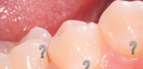
Caries in the most vulnerable areas of the teeth - in the area of fissures
Generally speaking, such nosologies as hidden caries are not in medical reference books and encyclopedias. Although the dentists themselves and their patients by this term ...
As the name suggests, the peculiarity of fissure caries is that it affects the so-called fissures - natural anatomical p ...
The term "internal caries" ordinary patient dental clinic usually understands the disease, affecting tissue deep under the tooth enamel. ...