

Compared with the carious lesion of enamel and dentin, cement caries or, otherwise, “subgingival caries” (root caries) is much less common, but unlike them it is a more aggressive and dangerous form for a tooth. Since the root of a tooth has a small wall thickness, its destruction by caries often takes place in a fairly short time, up to the development of pulpitis or periodontitis, which, in turn, sometimes even result in tooth extraction.
Since cement caries is often combined with cervical caries, for the front teeth, in addition to the risks mentioned, it is also fraught with aesthetics. Dark spots or carious cavities on the front teeth, especially if they are not eliminated for several years, often provoke psychological complexes, problems at work and in communication with the opposite sex.

To avoid all this, it is necessary, as they say,know the "enemy" in person: this is why already now it is possible and necessary to get clear and accessible information about how to recognize caries of cement in yourselves, what symptoms it can accompany and how to carry out the treatment with the maximum result to save the tooth. This and much more will be discussed further.
Risk factors for the development of caries cement
Most often (in about 60-90% of cases), tooth cement caries develops in older people due to gum diseases of various origins. In most cases, a pathological pocket is formed between the gum and the tooth - a place of accumulation of various microorganisms that not only provoke destruction of the periodontal attachment, which leads to tooth loosening, but also causes dissolution of the root cement with a depression in the root dentin (streptococcus).


On a note
According to the international classification of diseases, caries of cement goes after carious lesions of enamel and dentin, and is not often seen at a dentist’s appointment. The classification of carious cavities according to Black (Black) allows conditionally assigning caries of cement to class V - cervical defects of all groups of teeth.The conditionality is determined by the fact that the cervical defect is not always combined with the development caries under the gum; just as subgingival caries does not always extend beyond the borders of the gingival margin to the visible surface of the tooth.
The result of the destruction of cement and dentin caries is first the formation of a small carious cavity, which sooner or later leads to the penetration of infection into the tooth with the involvement of pulp (nerve) tissue in inflammation.
Additional risk factors leading to cement caries:
- Cervical or circular caries. If the carious process in the gum area gets access to the cement of the root of the tooth, then a kind of "double" caries is formed with two types of localization: above the gum and under the gum. It plays a role or a violation of the fit of the gums, covering the neck of the tooth, or denudation of the root for any reason.

- Incorrectly installed crown or violation of the statute of limitations for its fixation. If there are errors in prosthetics with crowns, it is possible that the edges of the gingiva will be unnecessarily introduced under the gum, or it may be insufficient to reach the gum boundaries established by norms. The result of this is either a gum injury with the formation of local gum disease,or a constant delay of food in a place where the crown does not reach the gingival margin, which also leads to inflammation. As a result, cariogenic microorganisms can easily penetrate under the gum with the involvement of root cement in the process.


- Violation of oral hygiene. Constant accumulation of plaque in the cervical area of a tooth or poor-quality crown without proper and regular hygiene often leads to gingival and subgingival caries due to cariogenic factors for dissolving tooth enamel and root cement.

Clinical signs
Depending on the location of the carious lesion under the gum, a clinic is typical of cement caries. So, when localizing caries in the periodontal pocket, when the inflamed gum closes the root from external irritants, it is a closed arrangement. In such cases, the root caries clinic is not bright. As a rule, a person has no pain or they are expressed slightly.
With an open arrangement of caries of cement, in addition to the root, the cervical area is also involved in the process of destruction. Depending on the depth of the carious lesion, there may be complaints about:
- violation of aesthetics (especially on the front teeth)
- eating discomfort
- the occurrence of pain from chemical (sweet, sour), thermal (cold and hot) and mechanical (when food penetrates under the gum) stimuli.

Feedback
Not so long ago, I had blackness near the gums near the upper tooth and it became sick. At first it was not even black, but some kind of brown stain, which I could not clean off with toothpaste, but then my gums began to bleed, and the stain began to grow every month. As a result, it hurt me to drink cold water and brush my teeth because of a sore gum. Since I work as a sales consultant, I have to communicate with people, and the front tooth with blackness catches my eye, all the more - I also became sick. The dentist said that this is already beginning caries of the root, which must be urgently treated until it has damaged the nerve. First, they removed the bloom and stone from all teeth, and after 3 days they put in a beautiful filling. Now nothing hurts.
Yaroslav, Reutov
Diagnostics of caries of cement, without leaving the house
With a closed arrangement of caries of cement, it can be very difficult to find a defect in oneself. In such cases, it is usually detected only during the procedure of curettage (curettage) of the pathological gum pockets, or during plastic surgery of the gums at a dentist-surgeon or a dentist-periodontist. Since the boundaries of the defect do not go beyond the edge of the gum, it is only when the pulpitis and pulpit pain occur that one can independently understand that this tooth has a hidden problem.

It is important to know
The acute form of pulpitis is characterized by severe spontaneous pain, which occurs even without external stimuli. Depending on the stage of inflammation of the "nerve" and the protective mechanisms of the body, the duration of pain is determined: from a few minutes to 1-2 hours. Most often, the pain intensifies in the evening and at night.
Chronic forms of pulpitis can develop, bypassing the acute stage, and manifest themselves as long aching pains, which can be aggravated by food stimuli (more often from hot). The chronic course of pulpitis can last up to 2-3 months or more, up to the transition or in the exacerbation of pulpitis with an acute clinic.spontaneous pain, or in periodontitis - inflammation of the tissues surrounding the root of the tooth, which often leads to its removal.
With an open arrangement of caries of cement on the front teeth in combination with cervical caries, as a rule, already at the stage of carious stain without a carious cavity and any symptoms, serious problems can be suspected and consult a doctor. Moreover, in this case we are talking about the comfort of communication with loved ones, friends, colleagues and other people. The appearance of dark dots, the cretaceous hue of enamel, its cracks and spalling at the border with the gum allows you to determine the caries of the cement at the initial stage of development, when it may be just “breaking through” into the subgingival region.

With extensive cavities, leaving the outer surface of the tooth deep into the gum, usually there are reactions to cold, hot, sweet, sour, as well as the sense of mouthwash, pain when eating. Often, the gum moves away from the tooth so much that under it is visible the caries-affected area of root cement and the root itself. In such cases, you must immediately contact a specialist to conduct additional studies and confirm the diagnosis.
Professional diagnostic methods
With a closed arrangement of caries of cement root, additional manipulations are required for making a diagnosis using instrumental and hardware methods. In the framework of the differential diagnosis can be applied the following approaches:
- Removal of supragingival and subgingival dental plaque: cleaning of plaque and calculus from all surfaces of teeth. Since gum diseases are most often provoked by tartar and plaque, in order to make a correct diagnosis, it is necessary to thoroughly clean the inspection zone from deposits. Manual methods (scalers, chisels, curettes, etc.), ultrasonic tips and devices for ultrasonic cleaning of teeth (tip for the dental unit Scaler, Piezon-master, etc.), as well as dental treatment with an Air Flow apparatus are used for this purpose.

- Careful isolation of the examined root from saliva. For this purpose, cofferdam is used - as the best option for protection against saliva and the convenience of examining the root, but you can also get along with regular cotton rollers.
 allows you to isolate the tooth from the rest of the oral cavity")
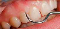
- Sounding root surfaces. It uses only a sharp probe, which allows to distinguish healthy tissue from caries affected by the characteristic surface roughness.

- Radiographic study.It allows not only to detect subgingival cavities in a suspicious tooth or under the crown, but also to reveal the slightest near-gingival defects in the region of the contact walls tightly adjacent to each other. At the same time, even a slight “darkening” can be seen on the X-ray of the tooth, which indicates that X-rays easily pass through the caries-affected tissue, which means that the carious process has already affected at least the cement, and at the most - dentin of the root. To detect a caries hidden under the gum, they widely use a viziograph, a device that transmits data to a computer and allows you to identify a defect and view it in an enlarged image or at a different angle.

The ideal option is a set of diagnostic measures combining the data obtained by the patient during self-diagnosis with a description of characteristic complaints, as well as the consistent application of professional diagnostic methods - from the removal of tartar and plaque from all surfaces of the teeth to x-ray diagnostics. This approach further allows a number of additional studies to be carried out. differential diagnosis of caries cement from pulpitis or periodontitis in case of difficulty. Namely: thermometry (the reaction of a tooth to cold water or a heated instrument), EDI (the reaction of the “nerve” of a tooth to a specific current, characteristic of a particular diagnosis, using electro-donometry), etc.
Modern approaches to treatment and the specificity of the choice of filling material
Modern approaches to the treatment of root caries allow the procedure to be carried out in one or several visits - this largely depends on the clinical situation. If the gum closes the carious cavity, bleeds, or is a serious obstacle to successful filling, then the gum correction (excision) is often carried out on the first visit.

After removing the interfering area of soft tissue, the carious cavity, after or without treatment, is covered with a temporary filling made of glass ionomer cement or regular oily dentin. After the healing of the gums of the patient is invited to re-admission and carry out filling.
The basic principles of the treatment of the cavity:
- Mandatory anesthesia, as root tissues are the most sensitive area for machining.
- Maximum excision of the modified and softened tissues on the root surface using modern techniques.
- Preservation of intact caries areas of the root surface.
- The formation of the cavity is rounded.
For the treatment of caries of cement used materials that are resistant to the influence of gingival fluid, saliva and blood during the filling of the tooth. Such materials are glass ionomer cements and compomers.


From the observations of the dentist
For patients who neglect oral hygiene, it is recommended to use glass ionomer cements that provide long-term fluoridation of tooth tissues after the filling. Most modern glass-ionomer materials have acceptable aesthetic characteristics that allow them, in certain clinical cases, to be installed even on the front teeth.
Light-cured composites can be used in combination with combined techniques, such as open sandwich techniques,when glass ionomer cement or compomer is first introduced and distributed in the subgingival cavity, and already in the subgingival region (in the smile zone), a composite seal with enhanced aesthetic qualities is simulated. Thus, the positive properties of each of the materials used are maximally used to achieve long-term fixation of the future filling, its strength and external perfection.
To control the quality of treatment, it is necessary to come to a second reception after the filling in 2-3 days (with artistic restoration) and, necessarily, after six months, to undergo a routine inspection to eliminate defects of the filling and caries recurrence.
How much can the treatment cost
As a rule, private clinics set prices for services based on the complexity of treatment and the cost of the materials used. In addition to the status of the clinic, its level of equipment, training, etc. treatment of caries of cement is laid in the price list as the most complex in technical implementation. At the same time, the price is fixed for the use of certain devices and preparations during treatment (for example, for excision of gums that have grown into the carious cavity), as well as materials for fillings: glass ionomer cements, compomers, composites, etc.

Combined techniques with the use of cofferdam to isolate the working area, working in 4 hands with a dental assistant, and treating caries for cement in 2-3 visits are, of course, more expensive than simple filling.
And the use of orthopedic dental treatments (crowns, inlays) with or without therapeutic measures (fillings) are several times more expensive.
An attempt to diagnose and treat root caries for free (according to the OMC) can end in tears - do not forget that this is a difficult case. Due to the workload and poor equipment of most dental offices (especially in rural areas) and clinics, there is a high risk of getting a free or cheap seal, which, if a violation of the filling technique occurs, will fall out in a few months. In the worst case, a wrong diagnosis by the doctor can lead to the occurrence of pulp pains already under the established filling, thus, it is possible to lose time for the tooth retreatment or even the tooth itself due to complications.
Dentist advice
For the treatment of caries of cement to be effective, you can choose any dentistry (even public),but it is important to find out from relatives, friends or acquaintances about the level of equipment at the clinic, reviews about specialists, approaches to treatment, long-term results, materials used, etc. If you want to save money, then you should be the last to think about comfort and service, since well-known companies charge up to 30-40% of the cost of treatment for this category of services.
Useful video about the caries and its characteristic features
About gum diseases and what they can cause (about periodontitis)
In most cases, tooth decay occurs in elderly patients over 60 years of age. This category takes about 60-90% of all cases of discern ...
Generally speaking, such nosologies as hidden caries are not in medical reference books and encyclopedias. Although the dentists themselves and their patients by this term ...
From the point of view of the causes and development of the pathological process, caries under the gum is no different from caries on the surfaces visible to the eye ...
Is it possible to treat caries of cement from the inside before setting a permanent filling (pulp is removed, channels are treated, temporary filling)?
Hello! If you mean treatment through the cavity of the tooth, where there is the possibility of deep penetration to the damaged wall, then sometimes this can be done, but there is a possibility of leaving carious areas on the outer surface of the tooth, where access is difficult. Ideally, the review of this part of the tooth should be favorable, better from the outside through the retraction of the gums in order to carry out a complete excision of carious tissues through this access. And already inside the "dead" tooth, this process can be controlled.