

Manifestations of caries are very diverse. Not only does this disease have several different forms, depending on the location on the teeth, it also divides the development of caries into several stages, at which carious lesions look quite specific.
For example, in the photo below - generalized caries:


And this photo shows a single tooth decay:

As you can see, the difference is quite significant and clearly demonstrates that caries can look very, very different.
The following shows caries at a very early stage of development, when it is a white or chalky spot (this is what the areas of demineralized enamel look like):

And in this photo - deep caries, which already threatens to turn into pulpitis:

It is interesting that at the stage of stains on caries most people do not pay any attention - you will think, the tooth slightly changed color, you never know what is wrong with it.And in vain, because it is at this stage that caries can still be cured without a drill, filling and violation of the integrity of the tissues of a living tooth. If you miss the moment, then without installing a seal (at best) it is hardly possible to get by.
In many cases, caries develops where it is very difficult to notice (hidden caries). Consequently, even if you know well how tooth caries look, you cannot be completely sure that if you don’t see it in your mouth in front of a mirror, it means that you don’t have it at all. In some cases, even a doctor with a special instrument may not notice carious damage, and sometimes a disease can be diagnosed only with the help of special dental examination methods (X-ray, transillumination, fluorescent diagnostics, etc.).
For example, the photo below shows an x-ray of a tooth with a hidden carious cavity:


If the enamel was damaged on the approximate surface of the tooth, it is extremely difficult to see such caries even with a dental mirror.
However, knowing how caries usually looks at different stages of its development, a person can detect damage to his teeth in time and consult a doctor.This often helps to save both teeth and a significant amount of money, so let's take a closer look at the full range of carious lesions closer ...
Types of caries: a visual demonstration
Caries is a disease that damages the hard tissues of the tooth: enamel and dentin. With an initial enamel lesion, the carious area usually becomes milky white and loses shine. When it comes to dentin, by this time the portions of the porous demineralized enamel, and locally the dentin itself usually looks brown or even almost black, as they are colored with various substances, including dyes from food.
Thus, the stage of the disease, the nature of its course and the localization of the lesion complement the picture and determine how exactly the caries looks on the teeth: color, size of damage, depth of the carious cavity, number of lesion areas, etc.
For example, in the photo below - fissure caries. On the teeth, it appears on the chewing surface in natural grooves, which are called fissures. In some cases, the black lines are so small that the dental probe and the doctor’s eyes do not linger on them, but often the problem is clearly visible to the naked eye:

Fissure caries is dangerous in that extensive patches of affected dentin can hide under seemingly insignificant damage to the enamel. For a better understanding, watch the video, which schematically shows how caries penetrates deep-seated tooth tissues:
Interesting information about the structure of the tooth and its caries
And this photo shows another example of how teeth may look affected. generalized caries. This is a very dangerous form of the disease that threatens a person with the loss of a significant part of the teeth, depulpation (removal of nerves) and the installation of crowns:

In appearance and character of flow, it is close to generalized bottle caries. The main difference between these diseases is in the age of the people they infect: bottle caries develops predominantly in children from 1 to 3 years old. Often this is due to a weakened immune system and somatic diseases of an early age.
Photos of bottle caries:


On the note about bottle caries
The causes of bottle caries go far beyond feeding a child with milk, especially before bedtime, as some parents think.It is important here: violation of the feeding regime, non-compliance of the parents with hygiene rules in relation to milk teeth, the factor of systemic diseases of the baby, aggravating the background of carious processes, directly and indirectly affecting the already formed and not yet fully mineralized enamel grid, the composition of the baby’s saliva is also important. In general, the definition of a bottle of milk - caries can not be limited.
A separate form is recurrent caries, appearing on previously cleaned surfaces. It usually develops at the site of poor quality tooth treatment around the filling. In the late stages of development, caries-affected tissues frame the filling and are clearly visible with the naked eye.
The photographs show how caries develops under the filling, manifesting itself in the places of its adhesion to the surrounding tooth tissues:


Another option for hidden caries is caries under the gum, where it hits the cement of the tooth root. For this reason, it is also called root caries or cement caries. It develops faster than caries of the open areas of enamel, but it is very difficult to see it.
The photo below shows the extracted tooth with root caries:

If the disease develops in the cervical area of the tooth, that is, in areas close to the gum, then in this case we are talking about cervical caries. Such a pathology also has a characteristic appearance: at the base, the tooth appears to be undermined, and sometimes along the entire perimeter, while the upper part of the crown often remains intact.
The following photographs show several examples of cervical caries:


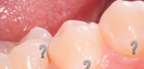
Proximal caries is very difficult to detect (sometimes it is called more simply - interdental caries): it develops on the surface of the tooth in contact with the wall of the adjacent tooth. Due to the difficulty in the complete cleaning of the interdental space with a toothbrush, such caries can long and quietly erode the enamel, and then dentin, often leading to the fact that a person turns for help at the stage of deep carious lesion of the tooth.
An example of approximate (interdental) caries - note that it is almost invisible:
 caries often proceeds in a latent form, visually without giving itself away.")
But in the case of a strong proliferation of the carious cavity between the teeth, such caries looks quite, so to speak, classically:

It is important to consider that,Although most of the above photos with caries of the teeth show the disease at sufficiently late stages of development, when the lesion and staining of the dentin began, this does not mean that caries is certainly brown or black spots and holes in the teeth. The color of the affected areas depends on the stage of development of the disease, and can vary greatly, ranging from snow white to black, including all sorts of variations of brown, gray and yellow.
Different stages of caries, different external manifestations
The photo shows caries at a very early stage of development:

This stage of development of the pathology is called the white spot stage: here only the initial damage of the enamel occurs and the formation of pores in it under the influence of aggressive acids from the oral cavity. Enamel loses shine, gets a lighter, more intense white color.
Here are some more photos of caries in the stains stage:


And then - the appearance of carious areas already after a through-hole had formed in the enamel and the dentin lesion began:

The defeat of the dentin marks the onset of the stage of average caries.
Next on the photo - caries under the microscope:

On a note
Dentin is much more porous and soft than enamel, and easily absorbs various coloring matter from the oral cavity. That is why after the start of the lesion, he quickly turns dark.
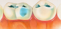
The picture shows the main stages in the development of dental caries:

Under the influence of cariogenic factors, dentin is destroyed much more easily than enamel, and therefore the development of medium caries and its transition to deep is usually quite rapid.
With deep caries, as the name implies, the deep layers of dentin up to the areas near the pulp are affected. With the defeat of the pulp itself, pulpitis begins with sharp pains and the risk of periodontal inflammation.
An example of deep caries on an x-ray is shown below:

Important!
Deep caries can develop even without visible lesions on the tooth surface. On X-ray, it is clearly visible, and therefore a snapshot is often taken to diagnose it and determine the extent of the lesion.
Depending on the place of manifestation, deep caries looks different. For example, in the photo below - a fairly significant damage to the tooth with caries:

And here - the teeth with deep carious cavities on the chewing surfaces:

X-ray caries: how it looks and why to photograph it
X-rays of carious teeth allow you to get the most complete picture of tissue damage without opening the enamel. The principle of radiography here lies in the fact that a beam of x-rays is passed through the tooth, which are attenuated in different ways when the tooth tissues pass through. When projected after such a passage on a sensitive sensor (or film), they demonstrate areas where there are damaged or no tissue at all - such areas look darker.
For example, in the X-ray picture below, a carious cavity in the tooth is clearly visible, which could not be detected by a simple visual examination:

X-rays are especially useful for detecting recurrence of caries, as well as root caries and approximate caries. For example, here is a photo where you can see what the approximate caries looks like in the picture:

What brings abstention from visits to the doctor
Due to delaying a visit to the dentist for too long, multiple caries can develop, which is unlikely to add to the attractiveness of someone:
..
Here is another photo showing the simultaneous destruction of several teeth at once by deep caries:

And here - acute caries on the front teeth of a child:

Do not forget that the initial caries in the staining stage is usually easily amenable to remineralizing therapy at the dentist and can be treated without preparation of the tooth with a drill. You can not say about the deep carious lesions, which can not only cause pain from various stimuli, but also turn into a serious complication - pulpitis, when urgent help is often required, as it is unbearable to endure such pain.
Dentist tells
Many people often wonder: can a dentist treat a tooth for himself? I authoritatively declare that such cases occur in practice if the dentist does not trust anyone except himself, or does not have enough money for these procedures, working in a small town or village.
I can say from personal experience that it is difficult to treat caries for myself.You can risk only if the working field is clearly visible and the cavity does not have a large area. In spite of this, some familiar doctors did not like this: they self-administered anesthesia, passed the channel during pulpitis of the front tooth and even with a shot, an incision was made with a “flux” to reduce the pain. Most often this was due to the inability to seek help from colleagues on holidays (or weekends), or the remote location of the nearest dental office. It sounds unbelievable, but it’s a fact: these dentists are extremely fearless people!
The picture shows what happens when caries reaches the pulp and causes it to become inflamed:

The following photographs show how some stages of tooth depulpation (nerve removal) look like:


The sooner you notice signs of caries and consult a doctor, the more effective and painless the treatment will be. Caries on the chewing and buccal surfaces of the teeth, and on the front teeth - and on the vestibular surface, in many cases, can be independently detected already in the early stages.
For example, this is what the front teeth with white spots look like in the photo - foci of initial caries:

And then in the photo - the mouth with teeth affected by fissure caries. One can see, though not large, but clearly visible dark lines on the molars. Installation of seals here is no longer to be avoided, but it is quite possible to prevent pulpitis from developing.

The beginning or the possibility of the appearance of caries in the near future may also produce tartar. The photo shows an example of such deposits on the inside of the front teeth:

In any case, when looking in the mirror, you must first pay attention to those parts of the teeth that stand out significantly from their neighboring colors. Whether it is too white, beige or, especially, brown - the fact that it differs from the color of intact healthy enamel can already be a sign of a tooth being damaged. And this is a good reason to go to the dentist. If, along with such a non-standard color, the tooth is also worried, even if not permanently, it should be treated as soon as possible.
Interesting video: an example of the treatment of deep caries with the preparation of a tooth with a drill
Is it possible to treat caries without pain and without a drill? ICON technology
Generally speaking, such nosologies as hidden caries are not in medical reference books and encyclopedias. Although the dentists themselves and their patients by this term ...
A caries marker is a special substance that when applied to caries-affected tissues (demineralized enamel, dentin) is fixed on ...
Decompensated form of caries is, in fact, an intensively developing caries, a pathological process that proceeds very actively in solid ...
Thank you so much for VERY useful information! Such as you around the world and more!
More x-rays with explanations. And the information is very interesting. Thank you very much.
Why do doctors not say anything during the examination? .. For example, what should be done and what should not be done so that caries does not appear.
Hello! The fact is that doctors do not have much time to "talk."It is clear that we are talking about the prevention of the disease, but look at the queues that accumulate in hospitals. And in the corridor someone will surely say: “That he has been chatting there for so long - we have been waiting for 2 hours already.” In private dentistry (not all), the doctor has time, as he records you to work out for about an hour or more. Therefore, in addition to the main time, the dentist has 10-15 minutes to talk with you about anything. Sometimes a private trader is not able to pay attention, since a patient with acute pain can “wedge in” in his main time, and the additional income is much more important than talking (for this private trader).
Briefly about the main thing: to prevent caries from appearing, it is enough to brush your teeth with toothpaste containing fluoride. The percentage of fluoride is best agreed with the dentist. Cleaning teeth only after eating (at least 3 times a day), and also do not forget about the gaps between the teeth: irrigators or dental floss.
If you want to talk to the dentist about the hygiene and protection of the teeth from caries, then the most non-employed and competent in this regard are dental hygienists and periodontists. Good luck to you!
Svyatoslav, my dentist said to me that no, I can't brush my teeth 3 times a day, “the enamel will be damaged.” He said that it is possible only 2 times. Moreover, I am young, and there are no problems.
Hello! This is from the category: you can not eat 6 times a day (depending on what, how much and how). In general, brushing your teeth is useful after eating - this is the norm. Moreover, modern standards recommend brushing your teeth with irrigation with a special irrigator to clean the interdental spaces.
What I would like to say in contrast to your doctor: there are toothpastes that I would not advise to clean and 1 time a day. For example, bleaching pastes are generally contraindicated for some people. At the same time, there are toothpastes with low abrasiveness, that is safe for enamel. For people with sensitive teeth, it is advisable to use such pastes in combination with a soft toothbrush.
Professional dentists emphasize that after eating you need to remove food debris and plaque to avoid caries and gum disease. I advise you to check with your dentist, which toothpaste is suitable for your enamel.When they limit themselves in brushing their teeth, then questions begin: what to treat first of all, caries or gums? ..
To poke some doctors with their filthy muzzle in this article! Year! For a whole year I went to different doctors, telling them about the pain in the tooth after chewing hard food - they said everything was all right. As a result, she had to wait because of them that the tooth began to ache after any food. I came again, and they tell me that everything is all right with the tooth! Did she almost force the doctor to drill a tooth, and what do you think was there? Completely destroyed the inside of the tooth, along with the root! If you have the same situation, go to X-ray, do not be as trusting as I am. And such doctors need to be fired, without the right to work in the medical field at all.
My teeth are falling, what should I do? I have been going to private dentistry for the 5th time, I spent a lot of money. They do not warn about anything until the teeth collapse completely. This is for you to come and spend the normal money again. Tell me a cheap way to avoid tooth decay. And how to protect the teeth, otherwise I am 21, and my teeth are already breaking. This is the 6th tooth 🙁
Hello! Not all private dentistry is “bred” for money, and not in all cases it is in a particular dentistry, even if it seems that you get more money out of you there specifically.
There are some simple but effective recommendations: 1. If you are not satisfied with the doctor’s tactics or the clinic, change the doctor or the clinic; 2. If you want to protect your remaining teeth from the effects of caries - do prevention.
For greater savings, you can carry out prevention in public dentistry, but the doctors who work in them often have a lot of work and very little time. As a result, quality may suffer, so the best option is a normal private clinic, where the doctor has plenty of time for your money.
Prevention in private owners who are most interested in, so that caries does not turn into complications and so that it does not appear on the teeth at all, is not cheap. The fact is that for the purity of the teeth requires regular removal of dental cameos and plaque: for example, the stone is removed every six months. The dentist will determine which spots are caries and which are pigmentation. Initial caries (in the staining stage) can be treated without a drill - ICON method (not the cheapest option).Dark fissures (pits on the molars) can be treated with an invasive and non-invasive method of sealing the fissures, although more often these foci of future serious caries after excision of the drill will be sealed with a light-cured material.
When all this is done, the best recommendation after proper preparation of the oral cavity for use is proper maintenance of dental health, namely, optimal hygiene. This includes: the selection of toothpaste with its alternation, brushes, irrigator for cleaning interdental spaces. The irrigator is a cool thing; many dentists consider an irrigator for home use to be the best way to prevent caries on the contact walls of teeth. I, as a practicing dentist, will say that caries most often develops on the walls, right next to the gum, where there is a constant collection food. The irrigator "knocks out" all the remnants of food.
Hygiene is shown every time after meals. Fluoridation of teeth is shown 2-3 times a year according to indications. For some people, dentistry is the same category of expenses as women's hairstyles and the beauty of nails - that is, a mandatory component of the normal course of life.So go ahead!
Everything is very interesting and detailed! Thanks for the extensive information with photos!
I really liked the doctors, very good, thank you.